Newsletter
Stay up to-date with the latest imaging, analysis and metrology news from Digital Surf.

A team of researchers from the Environmental Electron Microscopy Group at the Institute of Scientific Instruments of the Czech Academy of Sciences (Brno, Czech Republic) used dedicated electron microscopy methods for a more comprehensive analysis of quartz micro and nanoparticles.
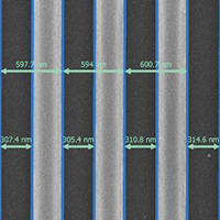
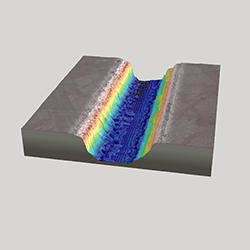
In the semiconductor industry, precise characterization of nanostructures is essential to ensure device performance and reliability. Line Edge Roughness (LER) and Line Width Roughness (LWR) play a critical role in process control. Mountains® software provides a dedicated solution for accurate, automated and robust LER measurements, ensuring reliable results regardless of imaging conditions such as accelerating voltage.
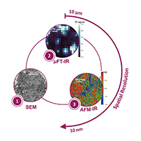
The AFM-IR lab at the Institute of Physical Chemistry, Paris-Saclay University (Orsay, France) is the pioneering research group in Infrared-Atomic Force Microscopy (AFM-IR). In particular, the team specializes in the characterization of complex materials, ranging from astro to biosciences, including studies on pathological calcifications such as breast microcalcifications (MCs), described in this article by Margaux Petay, former AFM-IR lab PhD student.
In the realm of dental implantology, understanding bacterial adhesion mechanisms is essential. Recent research led by Steve Papa and fellow researchers at the Jean Monnet University in Saint-Étienne, France, revealed the intricate relationship between surface topography and bacterial adhesion, with a particular focus on Porphyromonas gingivalis, a bacterium closely associated with dental implant failure.
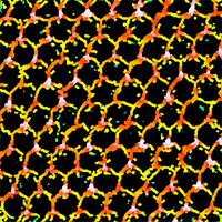
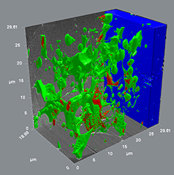
A team of researchers at the Catholic University of Louvain used topographic characterization and 3D reconstruction to reverse engineer the human ovary, bringing a plethora of new possibilities in biomedicine and biomimetics.
The Institute of Electronics, Microelectronics and Nanotechnology (IEMN) in France recently completed the “Dirac III-V” project investigating ways of producing Dirac electrons (electrons without any mass). This project called for the use of many different fabrication methods as well as a software program capable of bringing together and processing the different kinds of datasets generated.
A research team based at JEOL France recently studied the composition of magnetic fields incorporated in direct current electrical motors. In this study, they used a FIB-SEM technique coupled with a specialized analysis software package, based on Mountains® technology.
A group of researchers from the University of Ferrara, Italy, defined a general procedure to characterize surfaces and to evaluate their antimicrobial properties. Learn more about how they used Mountains® software in their study.
For this application, a research team at the LNE Nanotech Institute combined measurements from several instrument techniques including Atomic Force Microscopy (AFM) and Scanning Electron Microscope (SEM) equipped with a new-generation energy dispersive X-ray detector (EDX). They used MountainsLab® software to correlate the collected data and extract the relevant information.
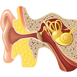
Incus bone erosion is considered a typical characteristic of advanced cholesteatomas (CHO), a pathology of the ear. Researchers at the Sapienza University of Rome explain how they used SEM image reconstruction technique to solve the mystery of this pathology and discover which cell erodes the middle ear incus bone.

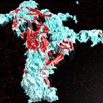
Bacterial infection of wounds is a major risk for patients undergoing skin grafts following severe burn injuries. Drs Monica Iliescu Nelea (left) & Michel Alain Danino, of the Plastic and Reconstructive Surgery Department at the University of Montreal Hospital Center (CHUM), Montreal, Canada are part of a group of researchers working on furthering medical understanding of this phenomenon.
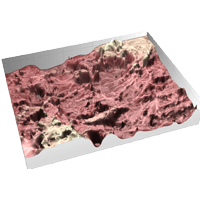
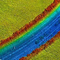
Ultrashort femtosecond lasers are known for their capacity to efficiently fabricate complex nanostructures and devices for a wide variety of applications. In two recent studies, the properties of femtosecond laser-structured surfaces were revealed thanks to a unique SEM image reconstruction technique.