In the realm of dental implantology, understanding bacterial adhesion mechanisms is essential to avoid peri-implantitis (a destructive inflammatory lesion of soft and hard tissues surrounding oral implants), and ensure implant longevity. Recent research led by Steve Papa and fellow researchers at the Jean Monnet University in Saint-Étienne, France, revealed the intricate relationship between surface topography and bacterial adhesion, with a particular focus on Porphyromonas gingivalis, a bacterium closely associated with dental implant failure.
Going beyond traditional roughness measurements
The study utilized femtosecond laser irradiation to create multiscale micro/nano-topographies, known as laser-induced periodic surface structures (LIPSS), on mirror-polished titanium samples.
These textured surfaces were meticulously analyzed using MountainsMap® software, offering unprecedented insight into surface properties. Instead of solely relying on traditional roughness measurements like Sa, the study explored a comprehensive range of parameters, including kurtosis (Sku), density of furrows and mean furrow depth.
MountainsMap® software was the central element in this research, providing a comprehensive platform for surface analysis. By exploiting the software’s capabilities, the researchers were able to go beyond conventional roughness measurements and gained a nuanced understanding of surface-bacteria dynamics.
The correlation analysis was carried out between each surface parameter and bacterial adhesion, allowing the precise identification of key topographic parameters in bacterial adhesion.
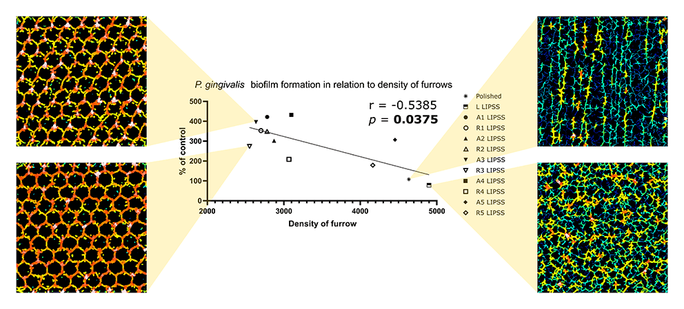
Above center. Graphical representation of the correlation between P. gingivalis adhesion and density of furrows or mean furrows depth with the Spearman’s rank-order correlation and associated p-value. Left & right. Qualitative rendering of the furrows.
Sku & furrow analysis
The software’s versatility allowed analysis of measurements at various scales, such as micro- to nano-topographies, providing a global view of surface characteristics.
The study underlined the limitations of conventional roughness parameters in capturing the complexity of surface-bacteria interactions. Instead, parameters like Sku and the aspect ratio between density of furrows and mean furrow depth were significant indicators of bacterial adhesion.
Surfaces with lower furrow density and greater mean furrow depth were found to promote higher bacterial adhesion, showcasing the critical role of surface complexity in microbial colonization.
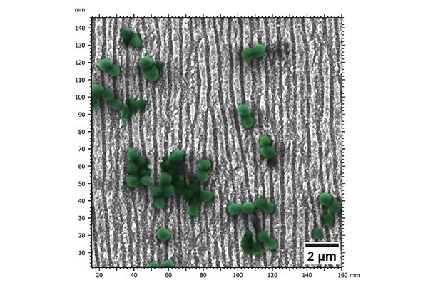
Above. Colorized SEM image of P. gingivalis on laser-induced periodic surface structures.
Furthermore, the research highlighted the potential for bacterial adhesion anticipation through surface evaluation, offering promising prospects for the field of ultrafast laser surface texturing. A more global and precise description of surface topographies would be instrumental in designing future biomaterials such as dental implants.
By revealing the intricacies of laser-textured surfaces with a great accuracy, MountainsMap® software is helping to pave the way to the development of next-generation dental implants with improved biocompatibility and longevity.
Above. 3D representation of laser-induced periodic surface structures.
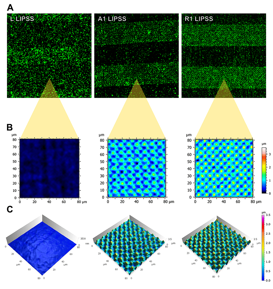
A. Representative fluorescence images of calcein-stained P. gingivalis after 48 h of contact on partially textured samples. B & C. 2D & 3D representation of surface topographies generated with MountainsMap® software.
Conclusion
This research represents a significant advancement in understanding the multifaceted dynamics of bacterial adhesion on implant surfaces. MountainsMap® software’s powerful and versatile tools enabled researchers to unlock new insights into surface/bacteria interactions, ultimately driving innovation in implantology as well as improving patient outcomes.
Instruments & software used
Focus variation microscope, scanning electron microscope & MountainsMap® software.
Read more
Key topographic parameters driving surface adhesion of Porphyromonas gingivalis
S. Papa, M. Maalouf, P. Claudel, X. Sedao, Y. Di Maio, H. Hamzeh-Cognasse, M. Thomas, A. Guignandon, V. Dumas. Sci Rep 13, Article no. 15893 (2023) doi.org/10.1038/s41598-023-42387-5
Work funded by public grant from the French National Research Agency (ANR) under the “France 2030” investment plan, ref. EUR MANUTECH SLEIGHT – ANR-17-EURE-0026