Newsletter
Stay up to-date with the latest imaging, analysis and metrology news from Digital Surf.
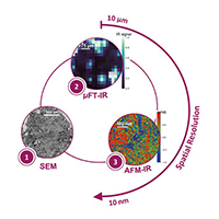
The AFM-IR lab at the Institute of Physical Chemistry, Paris-Saclay University (Orsay, France) is the pioneering research group in Infrared-Atomic Force Microscopy (AFM-IR). In particular, the team specializes in the characterization of complex materials, ranging from astro to biosciences, including studies on pathological calcifications such as breast microcalcifications (MCs), described in this article by Margaux Petay, former AFM-IR lab PhD student.
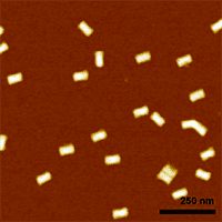
Simone Ruggeri, Associate Professor leading the Nanoscale Microscopy and Spectroscopy group at Wageningen University (WUR), Netherlands, tells us more about his current projects in the development of nano-analytical imaging and spectroscopic technologies to open a new research window of observation with nanoscale sensitivity in chemistry, biology and materials science.
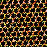
In the realm of dental implantology, understanding bacterial adhesion mechanisms is essential. Recent research led by Steve Papa and fellow researchers at the Jean Monnet University in Saint-Étienne, France, revealed the intricate relationship between surface topography and bacterial adhesion, with a particular focus on Porphyromonas gingivalis, a bacterium closely associated with dental implant failure.
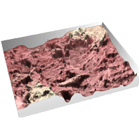
A team of researchers at the Catholic University of Louvain used topographic characterization and 3D reconstruction to reverse engineer the human ovary, bringing a plethora of new possibilities in biomedicine and biomimetics.
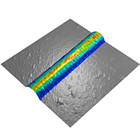
Researchers at the Structural Nanomechanics Lab at Dalhousie University in Canada have been investigating the nanomechanical behavior of Collagen I fibrils. Their study demonstrated that nanomechanical mapping can detect subtle changes in molecular dynamics and fibril architecture. This article explains how Mountains® software allows fine-tuning and detailed analysis of force volume data.
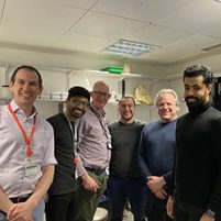
A team of researchers at King’s College London worked on ways to improve the understanding of the erosive tooth wear process, and also investigated innovative techniques for clinicians to monitor and treat it.

The profilometer manufacturer Nanovea conducted a study of different pharmaceutical tablets in order to study their surface roughness. With the use of a profilometer, they measured the average surface roughness of three different tablet surfaces. The data obtained was then analyzed with Nanovea’s Professional 3D software based on Mountains® technology.
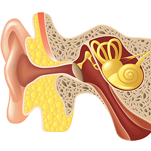
Incus bone erosion is considered a typical characteristic of advanced cholesteatomas (CHO), a pathology of the ear. Researchers at the Sapienza University of Rome explain how they used SEM image reconstruction technique to solve the mystery of this pathology and discover which cell erodes the middle ear incus bone.
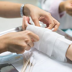
Bacterial infection of wounds is a major risk for patients undergoing skin grafts following severe burn injuries. Drs Monica Iliescu Nelea (left) & Michel Alain Danino, of the Plastic and Reconstructive Surgery Department at the University of Montreal Hospital Center (CHUM), Montreal, Canada are part of a group of researchers working on furthering medical understanding of this phenomenon.
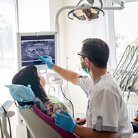
Researchers in tissue engineering & biophotonics at King’s College London (UK) seeking to attain better understanding of tooth enamel erosion recently applied its methods to bring to light micro-scale surface changes over time.

Bioceramics are particularly useful in the repair and reconstruction of bone. A group of researchers at the University of Limoges investigated the impact of bioceramic surface topography and composition on protein adhesion forces.
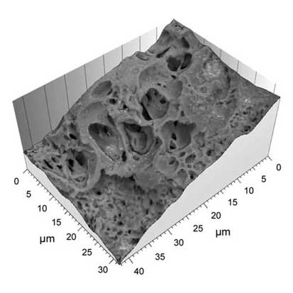
Using scanning electron microscopy and Hitachi map 3D software based on Mountains® technology, cell biology scientists at the University of Miyazaki (Japan) defined a new method for examining stem cell architecture.