Newsletter
Stay up to-date with the latest imaging, analysis and metrology news from Digital Surf.
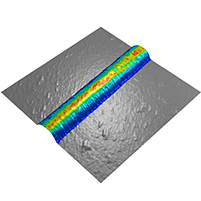
The semiconductor industry relies on uncompromised metrological precision. The optimal exploitation of AFM data requires powerful and adapted software, capable of transforming raw data into actionable insights. This is where Mountains® software comes into play, offering a high-performance solution for the precise and efficient analysis of Critical Dimensions (CDs) in the semiconductor field.
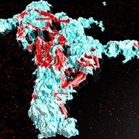
The AFM-IR lab at the Institute of Physical Chemistry, Paris-Saclay University (Orsay, France) is the pioneering research group in Infrared-Atomic Force Microscopy (AFM-IR). In particular, the team specializes in the characterization of complex materials, ranging from astro to biosciences, including studies on pathological calcifications such as breast microcalcifications (MCs), described in this article by Margaux Petay, former AFM-IR lab PhD student.
Simone Ruggeri, Associate Professor leading the Nanoscale Microscopy and Spectroscopy group at Wageningen University (WUR), Netherlands, tells us more about his current projects in the development of nano-analytical imaging and spectroscopic technologies to open a new research window of observation with nanoscale sensitivity in chemistry, biology and materials science.
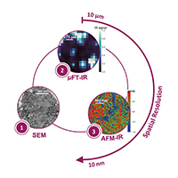
In the last few years, the use of SPM techniques in many areas of research has greatly increased. Professor Philippe Leclere, director of the LPNE at the University of Mons (UMONS) in Belgium, has been studying the impact of this increase and describes how his team is addressing the challenge of analyzing and mapping large quantities of data on material properties at the nanoscale.
Researchers at the Institut de Ciència de Materials de Barcelona characterizied surface texture of superconductor materials with the aim of improving their performance.
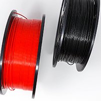
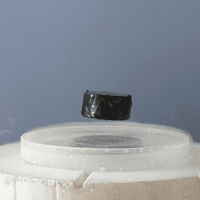
A team at Widener University (Chester, Pennsylvania, USA) recently used bimodal AFM technique and MountainsSPIP® software to study the calcification process of polylactic acid polymer (PLA), a plastic filament material widely used in 3D printing.
Researchers at the Structural Nanomechanics Lab at Dalhousie University in Canada have been investigating the nanomechanical behavior of Collagen I fibrils. Their study demonstrated that nanomechanical mapping can detect subtle changes in molecular dynamics and fibril architecture. This article explains how Mountains® software allows fine-tuning and detailed analysis of force volume data.
The Institute of Electronics, Microelectronics and Nanotechnology (IEMN) in France recently completed the “Dirac III-V” project investigating ways of producing Dirac electrons (electrons without any mass). This project called for the use of many different fabrication methods as well as a software program capable of bringing together and processing the different kinds of datasets generated.
For this application, a research team at the LNE Nanotech Institute combined measurements from several instrument techniques including Atomic Force Microscopy (AFM) and Scanning Electron Microscope (SEM) equipped with a new-generation energy dispersive X-ray detector (EDX). They used MountainsLab® software to correlate the collected data and extract the relevant information.
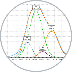
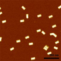
Scientists from the ISEN (Institut Supérieur de l’Électronique et du Numérique) in Lille, France, used photoelectron spectroscopy to investigate a new method for growing PbS nanoplatelets on InP surfaces.
Bruno Grandidier, research scientist with the French National Center for Scientific Research (CNRS), reports on his work, focused on understanding the electronic properties of silicon dangling bonds.

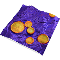
Bioceramics are particularly useful in the repair and reconstruction of bone. A group of researchers at the University of Limoges investigated the impact of bioceramic surface topography and composition on protein adhesion forces.