Dr. Mugino O. Kubo Ph.D is Lecturer at the Department of Natural Environmental Studies at the University of Tokyo, Japan. A long-time user of MountainsMap® software, Dr. Kubo speaks about her most recent research and how her team are implementing new, ground-breaking methods for studying the paleodiet of carnivorous dinosaurs, including the iconic Tyrannosaurus rex.

Please can you introduce yourself, your professional background, and how you came to be involved with Paleobiology, Evolutionary Biology and Ecology?
I’m a vertebrate morphologist with a strong interest in the evolutionary relationship between ecology and morphology. I started my career with a comparative osteological study using sika deer living in various environments in Japan. I have been focusing on how different feeding habits result in changes in dental and skeletal morphology. I collected and analyzed several hundreds of deer skulls. Based on such abundant skeletal specimens, I clarified the relationship between ecology and dental morphology, which can be used to infer the paleodiet of extinct animals. Since I married a dinosaur paleontologist, Dr. Tai Kubo currently at Okinawa Institute of Science and Technology, I got further involved in paleobiology. We have started a collaborative project on the paleodietary reconstruction of dinosaurs using dental wear traces.
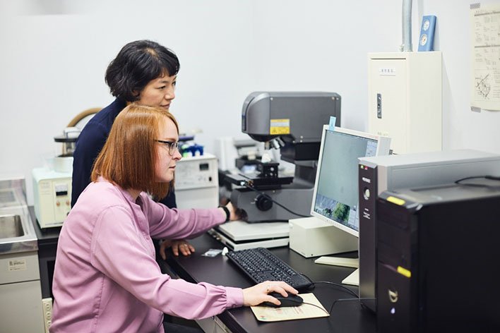
Dr. Daniela E. Winkler is scanning the surface of the tooth using a confocal laser microscope (VK-9700, Keyence).
What are your current projects?
I introduced a confocal laser microscope in my lab in 2016. This instrument can obtain 3D surface data at the sub-micron level. We used it to evaluate the roughness of tooth surfaces. When animals eat, tiny marks at the microscopic level (microwear) are left on the surface of their teeth due to contact with the food. As the microwear reflects the physical characteristics of the diet, investigating it allows us to infer what the animals might have eaten while they were alive. Since 2017, we have been working on research to accumulate microwear data for diverse wild animals with known diets, including sika deer (Fig. 1). Using these data as a comparison, we can reconstruct the ecologies of extinct species. We began our efforts with mammals and are now expanding to dinosaurs. Thanks to Dr. Daniela E. Winkler, who was a member of my lab from 2020 to 2022, we accelerated dental microwear research of dinosaurs. We published two papers on dinosaurs in 2022: on a Japanese sauropod, and on theropods, including Tyrannosaurus rex and its relatives (Fig. 2). We are now investigating hadrosaurs from various geological ages and extinct crocodiles from Cretaceous fossil localities.
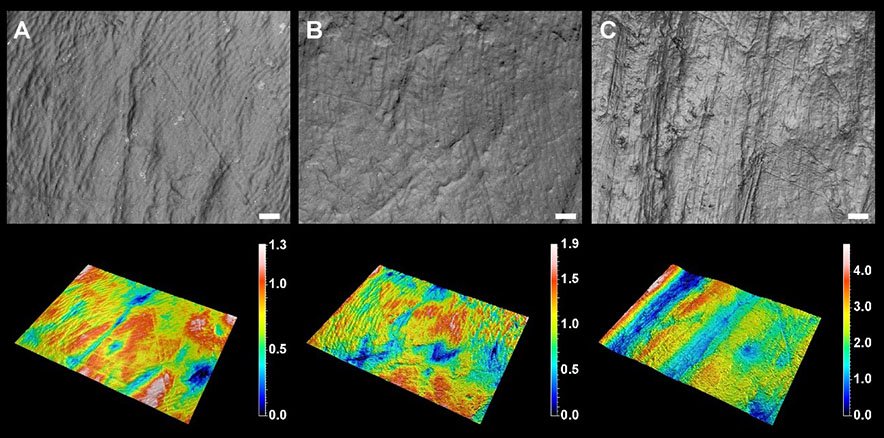
Fig. 1. 3D surface models of sika deer teeth generated with a confocal laser microscope. The field size is 140 × 105 µm. The height (color scale) is measured in µm. Among sika deer, individuals that eat more grass (C) have deeper wear marks than those that eat tree leaves (A and B). A: Yakushima Island, B: Shizuoka, C: Kinkazan Island. Credit: 2022 M. O. Kubo
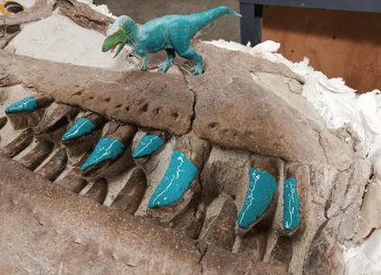
Fig. 2. Adult tyrannosaurid teeth (Teratophoneus curriei) at the Natural History Museum of Utah. Blue silicon was carefully excreted from a tube onto the teeth and left to dry for a few minutes to create near-perfect replicas, which were removed and taken from the museum in the U.S. city of Salt Lake City, Utah, to Japan for further study. Credit: 2022 D. E. Winkler
What is the innovation of your research and what is the most significant or exciting part to you?
Three-dimensional microwear analysis was proposed in 2004 by physical anthropologists and succeeded by mammalian morphologists and paleontologists. Currently, less than ten labs worldwide are actively conducting it. Each lab has its uniqueness in the scientific approach, and I would say our originality and novelty are in our focus on dinosaurs. To understand the diet-microwear relationship in carnivorous dinosaurs, we conducted controlled feeding experiments using live alligators, through an international collaboration with Professor Richard Blob at Clemson University in the U.S.A. Feeding experiments are usually only done with mammals, which are kept widely in laboratory environments: e.g., rats, guinea pigs, sheep, and pigs. But keeping and feeding alligators as a different challenge. Dr. Masaya Iijima, at that time Postdoc at Prof. Blob’s lab, fed individual alligators with different diets (crawfish, rats, quails, fish, and reptile pellets) and collected a lot of shed teeth. The teeth were sent to Japan and scanned by the confocal microscope to observe microwear differences. We were excited to see clear differences in wear marks, with higher surface roughness in more physically demanding diets (crawfish and rats) (Fig. 3).
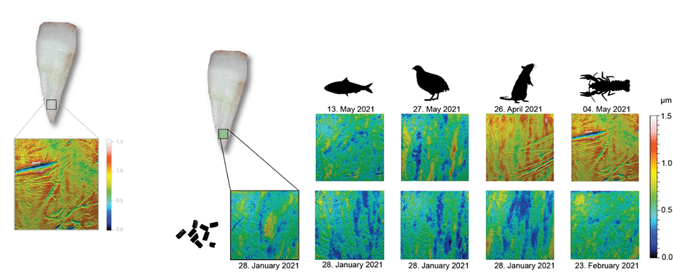
Fig. 3. Examples of microscopic wear patterns on alligator teeth. Each column shows teeth from the same individual, at different time points during the experiment. In the lower row, one tooth was collected while the alligators were feeding on pellets. Then, after about 3 months on their designated diets, rat- and crawfish-feeders developed distinct wear marks, while fish- and quail-feeders showed fewer wear marks. Credit: Winkler et al. (2022).
What role does MountainsMap® software play in your research?
MountainsMap® software plays an important role in our research projects. We use it from the initial stage of surface preparation through the calculation of surface roughness parameters defined by international standards (ISO 25178-2 and others). Our normal routine of obtained 3D surface data includes leveling, removal of gross tooth curvature, removal of measurement noises using spatial filters, and calculation of surface roughness parameters (Fig. 4). This procedure is quite similar to that used in Dr. Ellen Schulz-Kornas’s lab (Revealing diet from tooth wear surfaces in wild chimpanzees), but we used different spatial filters that can more effectively remove the measurement noise which is specific to our machine.
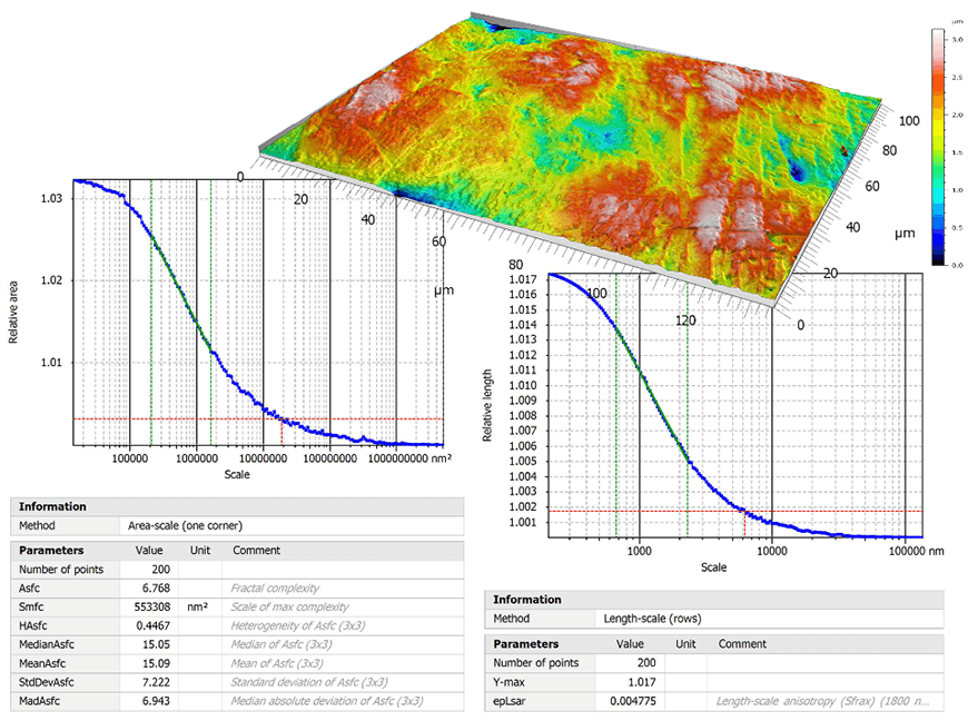
Fig. 4 Surface texture parameters including Asfc (area-scale fractal complexity), HAsfc (heterogeneity of complexity) and epLsar (exact proportion of length-scale analysis) calculated on the tooth surface of an African antelope (the hartebeest). Scale-sensitive fractal analysis can summarize the surface characteristics in a fewer number of parameters than ISO-25178 defined surface roughness.
Where can readers find more information?
You can find our lab page here (sorry but English version is under construction).
https://sites.google.com/edu.k.u-tokyo.ac.jp/mugino-kubo-lab/home
Press releases made in 2022 in English.
https://www.k.u-tokyo.ac.jp/en/information/category/press/9831.html
https://www.u-tokyo.ac.jp/focus/en/press/z0508_00264.html
Our latest selected papers on dental microwear texture analysis are available online (all free access).
Reconstructing diets of hunted sika deer from Torihama Shell Midden site (ca. 6,000 years ago) by dental microwear texture analysis
https://www.frontiersin.org/articles/10.3389/fevo.2022.957038/full
Microwear textures associated with experimental near-natural diets suggest that seeds and hard insect body parts cause high enamel surface complexity in small mammals
https://www.frontiersin.org/articles/10.3389/fevo.2022.957427/full
The dental microwear texture of wild boars from Japan reflects inter- and intra-populational feeding preferences
https://www.frontiersin.org/articles/10.3389/fevo.2022.957646/full
Controlled feeding experiments with juvenile alligators reveal microscopic dental wear texture patterns associated with hard-object feeding
https://www.frontiersin.org/articles/10.3389/fevo.2022.957725/full
First application of dental microwear texture analysis to infer theropod feeding ecology
https://onlinelibrary.wiley.com/doi/10.1111/pala.12632
T-rex image attribution 2.0 Generic (CC BY 2.0): Steve Jurvetson flickr.com/photos/jurvetson/11847683823
Instruments & software used
Keyence confocal laser microscope + MountainsMap® software (Scale-sensitive fractal analysis)